On your first Monday in an eye clinic, someone will ask you to fetch the “Goldmann” or check that the “indirect” is charged. If you haven’t heard those words before, you’ll nod and hope. This guide gives you ophthalmology instruments with names, plain descriptions and a sense of where each one lives in the clinic.
Think of it as a working eye examination equipment list rather than a textbook chapter. Some items sit in the consulting room all day. Others come out only for theatre lists or specific tests. Good new eye clinic staff training should cover all of them, but it rarely does so in one tidy place.
Two habits will help from day one. Learn the instruments by the order a patient meets them, and learn who is responsible for cleaning each one. The rest follows.
Diagnostic Ophthalmology Instruments: What Happens Before the Doctor Looks
Most patients meet the diagnostic kit before they meet the ophthalmologist. The chart projector or screen tests visual acuity, and the autorefractor gives an automated estimate of the glasses prescription. A lensmeter (also called a focimeter) reads the power of the glasses the patient already wears. These are quick, and they set the tone for the visit.
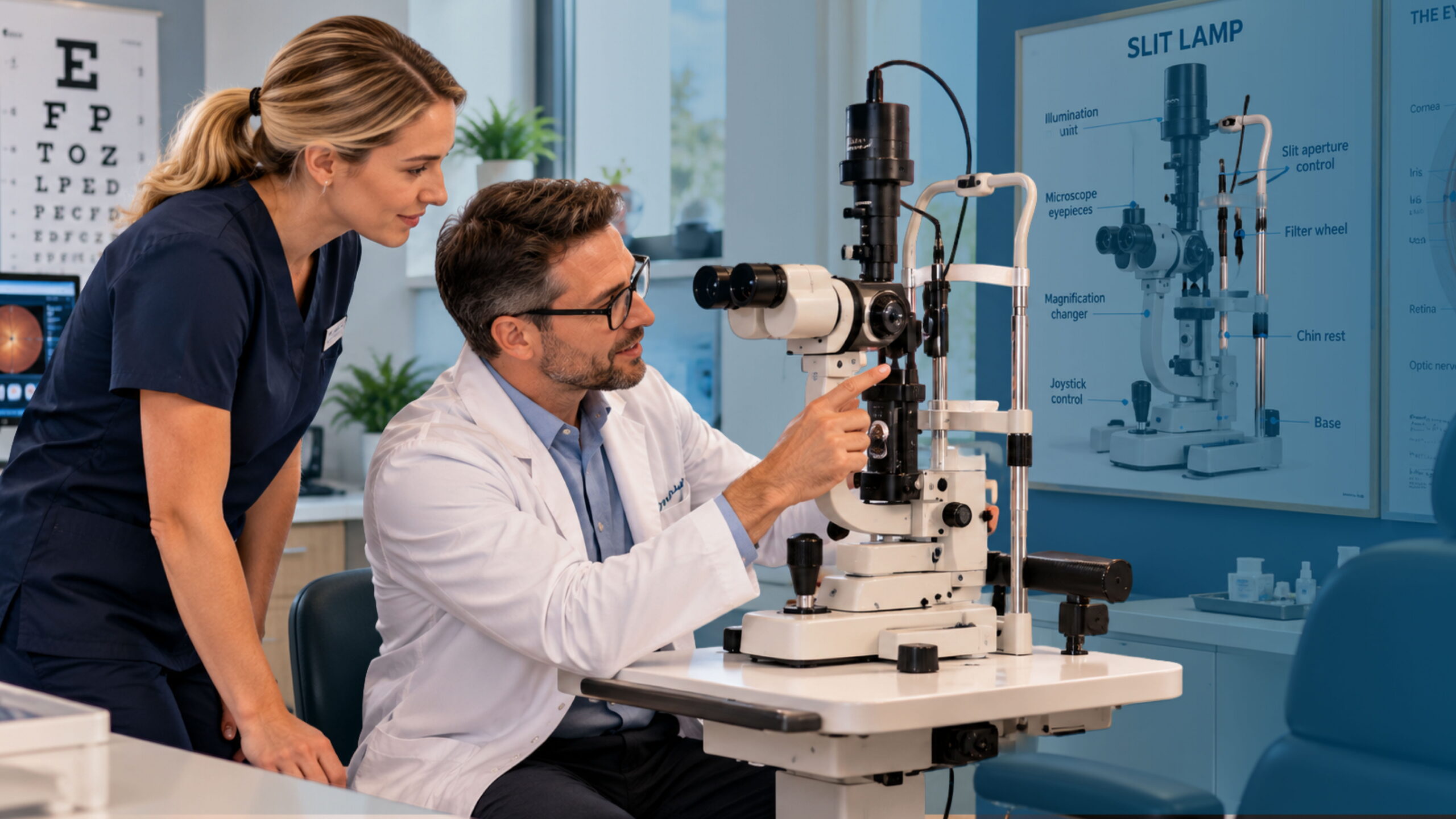
Then comes the pairing everyone talks about: the slit lamp and tonometer. The slit lamp is a table-mounted microscope with an adjustable beam of light. It lets the clinician inspect the eyelids, cornea, lens and, with a handheld lens, the back of the eye. The tonometer measures intraocular pressure, which matters enormously in glaucoma. The Goldmann applanation tonometer usually attaches to the slit lamp, while non-contact “air puff” devices are common for screening.
Further along the pathway you’ll find the ophthalmoscope, direct or indirect, for viewing the retina. A keratometer measures corneal curvature. A pachymeter measures corneal thickness, which helps interpret pressure readings. Optical coherence tomography (OCT) scanners produce cross-sectional images of the retina, and visual field analysers map peripheral vision. The American Academy of Ophthalmology’s practice management resources are a good place to see how clinics organise this equipment around real workflows.
A small detail that trips up newcomers: many of these devices produce images and numbers that must land in the right patient’s record. A pressure reading typed against the wrong name is a clinical risk, not an admin slip. Ophthalmology Times has described how modern records integrate imaging such as OCT and visual fields, and that integration is worth understanding early.
Surgical Ophthalmology Instruments and Their Uses
Theatre is where the vocabulary gets sharper, literally. Surgical ophthalmology instruments and their uses vary by procedure, but a few names come up constantly. If you assist in a day-surgery list, you’ll hear them within the first hour.
The eyelid speculum holds the lids open so the surgeon has a clear field. Forceps come in many forms: fine-toothed Castroviejo or Colibri types for handling delicate tissue, and tying forceps for sutures. Castroviejo scissors and Vannas scissors make tiny, precise cuts. A needle holder grips the curved needles used for closing wounds, and a lacrimal cannula is used to irrigate tear ducts.
Cataract surgery, the most common eye operation worldwide, relies on a phacoemulsification machine. It breaks up the clouded lens with ultrasound and aspirates the fragments. Alongside it you’ll see keratomes and blades for creating small incisions, a cystotome for opening the lens capsule, and an injector for placing the artificial lens. The World Health Organization’s page on vision impairment and blindness explains why cataract and other eye conditions drive so much demand for this work.
Instrument trays are usually set up as procedure-specific packs. Learn what a standard cataract pack contains, and you’ll spot a missing item before the surgeon does. That’s a quiet win nobody applauds, but the theatre nurse will remember it.
Not every clinic does surgery. If yours refers patients out, you still need the vocabulary, because referral letters and operation bookings mention these instruments and procedures by name. Understanding the terms makes you far better at scheduling the right theatre time.
Eye Clinic Instrument Cleaning and Sterilisation
Eye clinic instrument cleaning and sterilisation deserves more attention than most induction days give it. The eye is unforgiving. Residue on a tonometer tip or a contaminated surgical blade can cause real harm, and some outbreaks of eye infection have been traced to poorly processed equipment.
Start with a simple distinction. Surfaces and instruments that touch intact skin, such as chin rests and forehead bars, need regular cleaning and disinfection between patients. Instruments that touch the eye itself, such as applanation tonometer tips, need disinfection according to the manufacturer’s instructions, or single-use tips where your policy requires them. Surgical instruments that enter the eye need full sterilisation, normally in a steam autoclave after thorough manual or ultrasonic cleaning.
Here’s the part people skip: cleaning comes before sterilising. Debris and viscoelastic left in a cannula can shield microbes from the steam. Many clinics also keep a sterilisation log recording each cycle, the load and who ran it, so a problem can be traced.
Rules differ between countries and sometimes between regions, so check your local regulator and your instrument manufacturers’ guidance rather than assuming one standard applies everywhere. Professional bodies such as The Royal College of Ophthalmologists publish standards and guidance that many clinics use as a reference point, though your own jurisdiction’s rules come first.
A practical routine for new staff:
- Check the tray or device label and the date before every use.
- Never return an unclean item to a sterile pack.
- Report any dropped, damaged or doubtful item immediately.
That short list covers most of what goes wrong in practice.
Where GoodX Eye Fits Into Daily Clinic Life
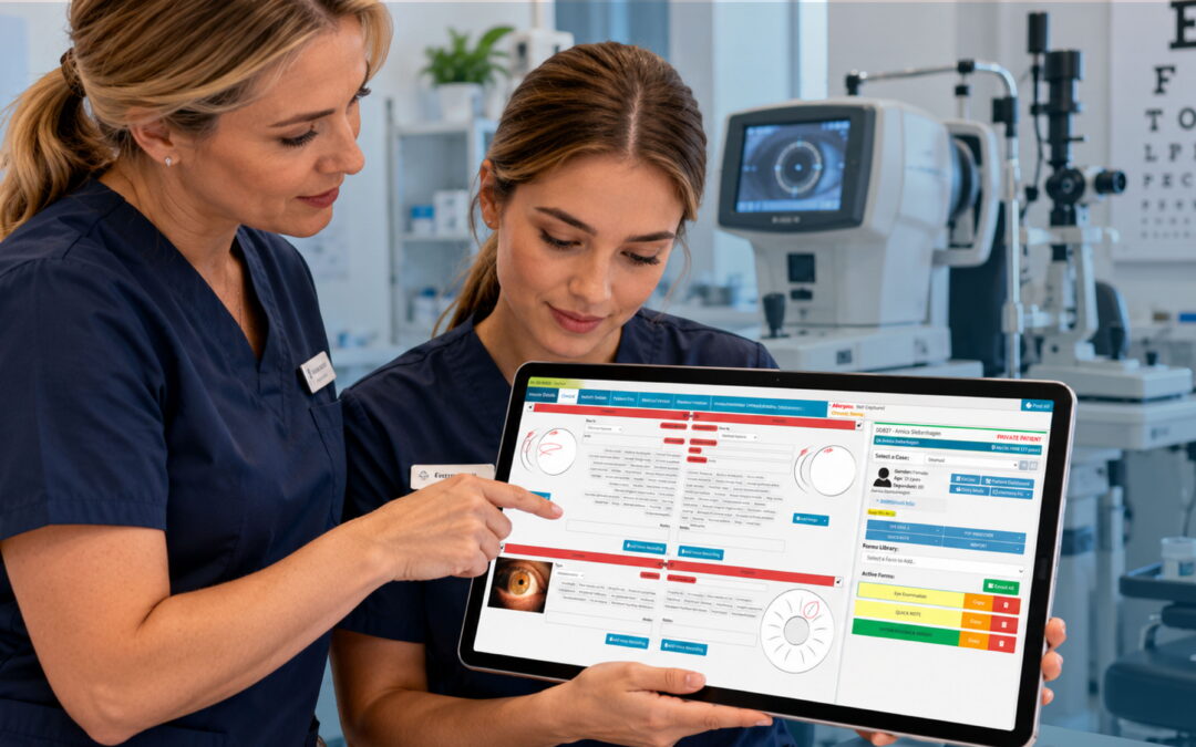
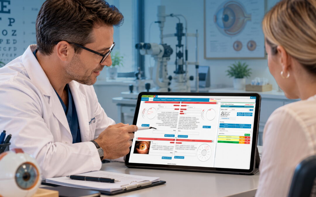
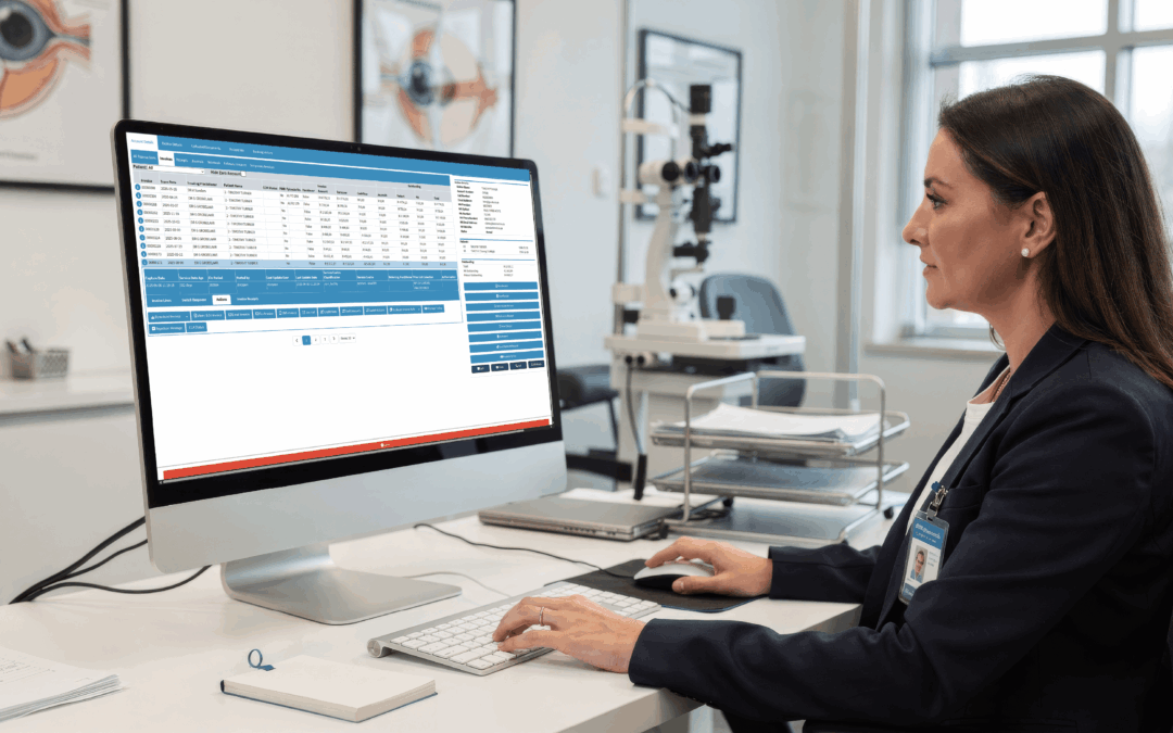
Instruments are only half the story. Every test generates a result, every result needs a home, and every visit needs a booking, a note and eventually a claim or invoice. GoodX Eye is practice management software built for ophthalmology and eye care practices, and it helps tie these threads together.
In a busy clinic, the clinical record is where diagnostic findings should end up, alongside the patient’s history. Clear records support continuity of care, which the Review of Ophthalmology has highlighted in its piece on keeping in touch with patients. Automated reminders and recall tools, such as those GoodX Eye supports for appointments, can cut missed visits so your diagnostic suite isn’t sitting idle on a Tuesday afternoon. Related reading: appointment reminders and recall.
Flow matters too. Seeing patients in arrival order and keeping them in one room while the clinician moves, as described in Better Ways to Manage Patient Flow, depends on staff knowing where each patient is. Scheduling and room status tools help here, and eye clinic scheduling software shows how that can work in practice.
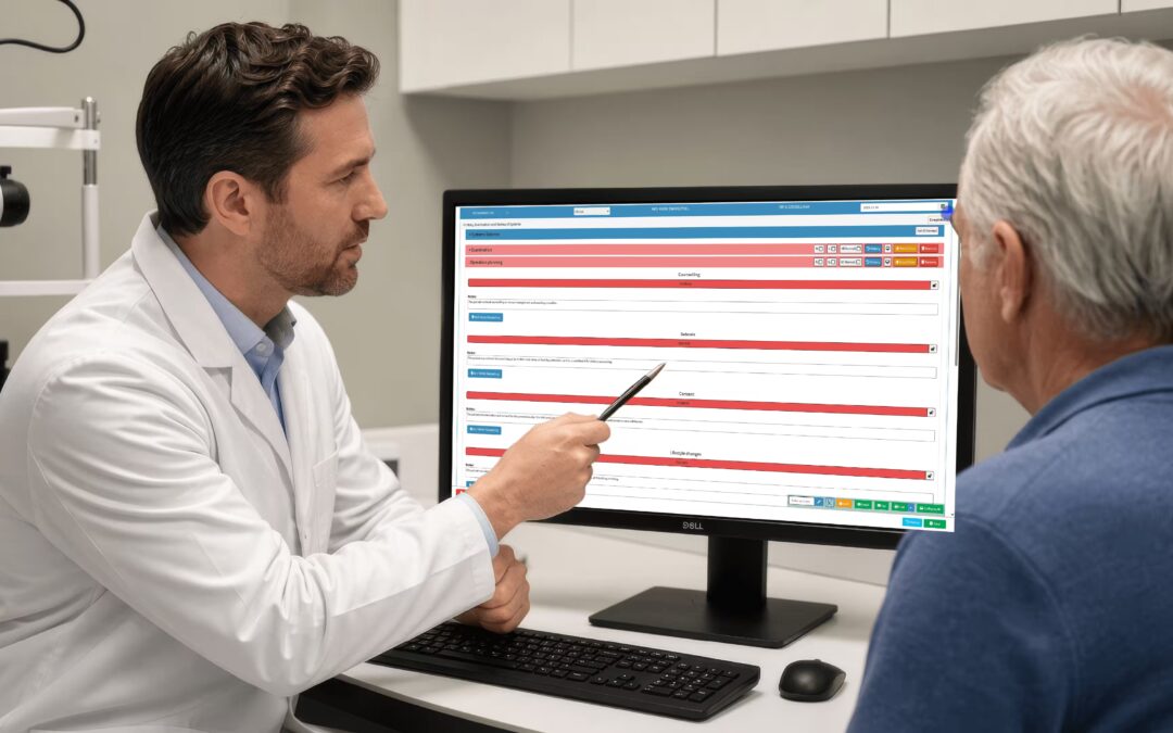
Behind the scenes, GoodX Eye supports billing and claims, reporting and record keeping, which help practices stay organised as they meet their local compliance obligations. For a new team member, a system that links bookings, records and billing means fewer sticky notes and fewer questions about where a result went. Related reading: eye clinic billing and reporting.
Frequently Asked Questions
What is the most important instrument for new eye clinic staff to learn first?
The slit lamp is the best starting point. It’s used in nearly every consultation, and learning its parts, such as the chin rest, joystick and beam controls, teaches you how patients are positioned. From there, the tonometer and ophthalmoscope are natural next steps.
What is the difference between a tonometer and a pachymeter?
A tonometer measures the pressure inside the eye, which is key for glaucoma care. A pachymeter measures corneal thickness. Clinicians often use both, because corneal thickness can affect how a pressure reading should be interpreted.
How often should eye clinic instruments be sterilised?
Surgical instruments that enter the eye need sterilising after every use. Diagnostic items that touch the eye need disinfection between patients, following the manufacturer’s instructions and your local rules. Always follow your clinic’s written protocol, as requirements vary by country.
Do receptionists and administrators need to know instrument names?
Yes, at least the common ones. Knowing the difference between an OCT scan and a visual field test helps you book the right appointment length, prepare patients and explain waiting times accurately.
Where can I find reliable training material for eye clinic staff?
Start with your clinic’s own induction and the manufacturer’s manuals. Professional bodies such as the American Academy of Ophthalmology and the National Eye Institute publish useful educational material for eye care teams.
Book your free GoodX Eye demo
If you’d like to see how GoodX Eye can support your team from the first appointment to the final claim, book a free demo. Visit goodxeye.com and we’ll show you how it works in a real eye care practice.
Disclaimer: This article is provided for general informational and educational purposes only. While GoodX Software takes reasonable care to ensure that the information is accurate and current at the time of publication, laws, regulations, industry standards, healthcare policies and technology may change. The content should not be regarded as medical, legal, financial or other professional advice. Readers should verify information relevant to their circumstances and consult an appropriately qualified professional where necessary. GoodX Software accepts no responsibility for decisions made or actions taken solely on the basis of this content.