by HannesE | Jun 26, 2026 | Blogs
Ophthalmology has been at the front of the AI in healthcare story from the start, and for a simple reason: the eye produces images, and images are exactly what these models read best. Diabetic retinopathy screening was among the first places an algorithm earned regulatory clearance to support real clinical decisions.
That head start cuts both ways. More promise, and more lessons already learned the hard way.
Here is a grounded look for eye practices: the genuine pros and cons, a straight answer on which country leads in AI, and the disadvantages worth weighing before a tool goes anywhere near a patient.
AI in Healthcare Pros and Cons
The upside in ophthalmology is unusually tangible. AI can screen retinal images at scale, flagging signs of diabetic retinopathy, glaucoma, or macular degeneration faster than a clinic could manually, which matters when screening volumes keep climbing.
The cons are equally specific. A model can miss an atypical presentation or flag a false positive that sends a worried patient for tests they did not need. Practitioners sometimes ask which medical jobs will be replaced by AI, and in ophthalmology the realistic answer is that screening throughput changes while the specialist’s diagnostic and surgical role does not.
The professional consensus is clear on where responsibility sits. The American Academy of Ophthalmology stresses that AI tools support, rather than supplant, the ophthalmologist’s judgment, and that a clinician reviews and owns the final read.
Which Country Is No. 1 in AI?
The honest answer depends on the yardstick. By research, investment, and the concentration of leading firms, the United States leads, with China close behind and ahead in certain applied areas, including some medical imaging work.
For ophthalmology specifically, leadership is less about geography and more about which regulators have cleared which tools, and on what evidence. A screening algorithm validated on one population may need fresh validation before it is safe on another.
For an eye practice, the practical question is narrow and local: is this tool cleared, validated on patients like mine, and supported. Research published in The Lancet keeps reinforcing that real world clinical performance, not national ranking, decides whether an ophthalmic AI tool actually helps.
Disadvantages of AI in Healthcare
The disadvantages take a particular shape in eye care. Image quality is the first. A poorly captured retinal photograph produces an unreliable result, so the tool is only as good as the camera and the technician behind it.
Then the familiar concerns. Bias, when a model was trained largely on one population. The black box problem, when a tool grades an image without explaining why. Over reliance, when a clinic starts trusting the screen over a second look. And patient data security, which carries real legal weight wherever you practise.
Sight is not a forgiving area to get wrong. The World Health Organization has documented the scale of preventable vision loss worldwide, which is exactly why AI screening is promising and exactly why it must be deployed with a specialist firmly in the loop.
Building AI Screening Into Your Clinic
For an eye practice, the practical question is not whether AI screening works in studies. It clearly does on narrow tasks. The question is how it fits a real clinic without creating new problems.
Image quality is where it lives or dies. A screening model fed blurry or poorly centred retinal photographs will produce results you cannot trust, so the camera, the lighting, and the technician’s training matter as much as the algorithm. Get the capture right and the tool is worth having. Get it wrong and you are reviewing noise.
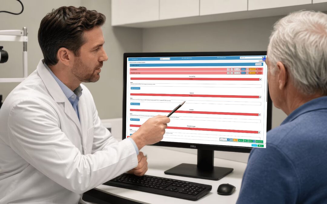
Build it in as a triage aid, not a verdict. Let the model sort the obvious normals from the cases that need a closer specialist look, and keep an ophthalmologist signing off every read that matters. Store the images and results in the same system as the rest of the patient record, so a flagged scan today connects cleanly to the follow up in three months.
Beyond Screening: Other Uses in Eye Care
Retinal screening gets the headlines, but it is not the only place AI is finding a role in ophthalmology. The eye generates a lot of data, and data is what these tools work on.
Models are being explored to track disease progression over time, comparing today’s images against a patient’s own history to spot subtle change a single visit might miss. Others support surgical planning, or help triage referrals so the urgent cases reach a specialist sooner. On the administrative side, the same automation that helps any practice applies here too, from scheduling to coding.
The thread running through all of it is the same. These are aids to a specialist, not replacements for one. The ophthalmologist’s judgment, examination, and surgical skill remain the core, with AI handling the volume and the pattern spotting around the edges.
Keep the Specialist in Charge
For all its promise in eye care, AI works best as a second set of eyes, never the final one. The ophthalmologist stays the decision maker.
The sensible model is simple. The model flags and sorts, the specialist reviews and decides. That keeps the speed of automation without surrendering the judgment that a difficult case demands. It also keeps responsibility where it belongs, with the clinician who can examine the patient, weigh the history, and act on more than a single image. Used that way, AI extends a specialist’s reach rather than competing with it.
Frequently Asked Questions
What are the pros and cons of AI in ophthalmology?
The pros are fast, scalable retinal screening for conditions like diabetic retinopathy and glaucoma. The cons include missed atypical cases, false positives, and dependence on image quality. Used with a specialist reviewing every read, the benefits hold while the risks stay controlled.
Which country is number one in AI?
By research, investment, and leading firms, the United States ranks first, with China close behind and ahead in some applied imaging work. For ophthalmology, what matters more is whether a specific tool is cleared and validated on patients like yours.
What are the disadvantages of AI in eye care?
Dependence on image quality, bias when models are trained on narrow populations, the black box problem where grading cannot be explained, over reliance by clinics, and patient data security duties. Sight is unforgiving, so a specialist must stay in the loop.
Will AI replace ophthalmologists?
No. AI changes screening throughput by handling high volumes of images, but it cannot replace diagnostic judgment, surgical skill, or the clinician who owns the final read. The realistic future is ophthalmologists working with AI as a screening aid, not a substitute.
Book Your Free GoodX Demo
AI screening works best when imaging, records, scheduling, and billing live in one connected system. That is exactly what GoodX is built to provide for eye practices.
See how a connected practice system would fit your clinic.
by HannesE | Jun 26, 2026 | Blogs
Billing for medical services in ophthalmology has a complexity all its own. A single clinic day can mix routine consultations, diagnostic imaging, minor procedures, and surgical follow ups, each with its own codes and rules. Get the billing right and the practice thrives. Get it wrong and revenue slips away one denied claim at a time.
Few ophthalmologists trained for this side of the work, and it shows in the rejection rates.
This guide breaks it down plainly: what medical billing is, what it looks like with eye care examples, the types of billing in play, and how the process runs from the appointment to the payment.
What is Medical Billing?
Medical billing is the process of submitting and following up on claims with payers so a practice is paid for the care it delivered. It works hand in hand with coding, which translates the encounter into standardised codes, turning a clinic visit into a paid invoice.
In ophthalmology that coding is unusually detailed. Procedures, laterality (which eye), diagnostic tests, and follow up periods all have to be captured precisely, because a missing detail is a fast route to a denial.
The diagnostic codes rest on a shared international standard. The ICD system maintained by the World Health Organization underpins the diagnosis side, and accuracy there is what keeps a claim clean on its first pass.
Medical Billing and Coding Examples
Eye care examples show why the detail matters. A patient presents for a cataract assessment. The encounter is coded with the appropriate examination code and the diagnosis, and any imaging such as biometry is captured separately.
Take a procedure. An intravitreal injection in the right eye needs the procedure code, the correct laterality, and often a modifier to distinguish it from a bilateral or repeat treatment. Miss the laterality and the claim bounces. Capture it cleanly and it pays without a fight.
This level of specificity is normal in ophthalmology, not exceptional. Coding guidance from the American Academy of Ophthalmology is detailed precisely because small errors in eye care claims are both common and costly.
Types of Medical Billing
Billing divides along a few lines that shape how an eye practice runs its revenue. The core split is professional billing, for the ophthalmologist’s services, versus facility billing where a procedure is performed in a surgical centre.
There is also the in house versus outsourced decision. A busy surgical practice might keep billing internal for tighter control over complex claims. A smaller clinic might outsource to a specialist eye care billing service and trade margin for less administrative burden.
Whatever the model, the medical billing process follows the same path: verify eligibility, capture charges, code the encounter, scrub and submit the claim, post payments, and work denials. Reviews summarised in The BMJ link well organised billing administration to stronger practice finances and fewer write offs, which in a procedure heavy field adds up quickly.
Where Eye Care Claims Leak Money
Ophthalmology billing leaks in places general practice never sees, and the leaks are specific enough that you can plug most of them deliberately.
Laterality is the classic one. A claim that does not specify which eye, or specifies the wrong one, bounces straight back. Bundling rules trip up practices too, when a diagnostic test billed alongside a procedure is denied as included. Then there are the follow up periods after surgery, where a visit billed inside the global period gets rejected because it should not have been charged at all.
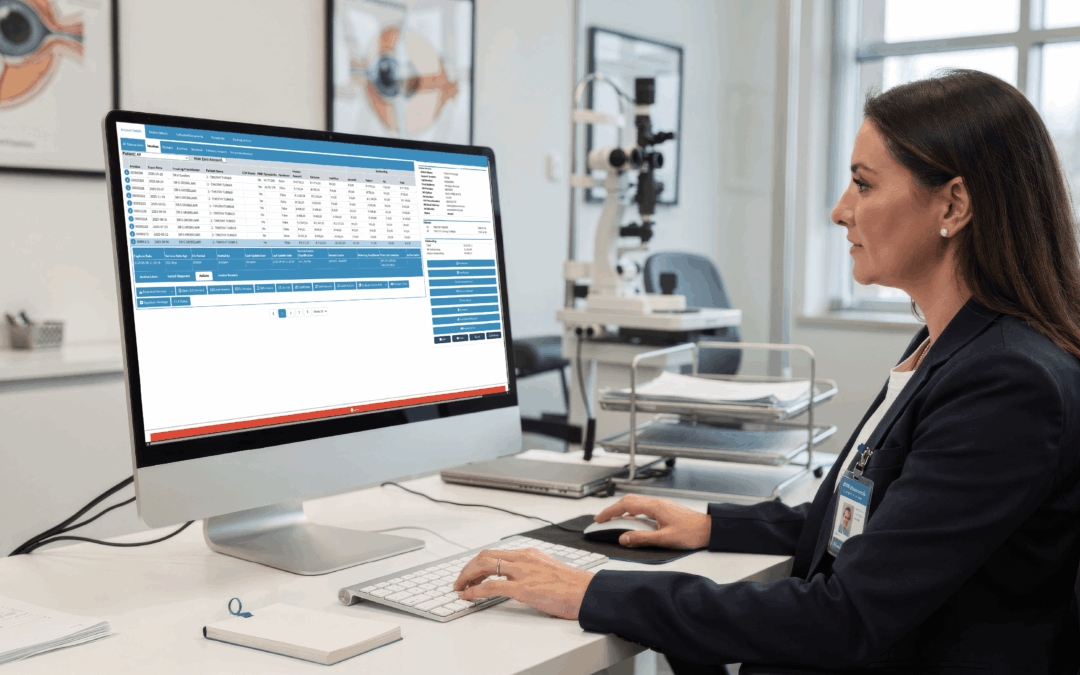
Software that understands eye care coding, not just generic medical billing, catches these before submission. It prompts for laterality, knows the bundling and global period rules, and ties the claim to the documented procedure. In a field where a single clinic day can generate dozens of coded items, that specificity is the difference between getting paid promptly and writing off claims you earned.
Getting Paid for Diagnostic Imaging
Ophthalmology runs on diagnostics, and imaging is where a lot of billing value sits, and a lot of it slips away through avoidable errors.
Tests like optical coherence tomography, visual fields, and fundus photography each carry their own codes and their own rules about when they can be billed and how often. Bill a test too soon after the last one, or alongside a procedure it is considered part of, and the claim is denied. Multiply that across a busy imaging schedule and the lost revenue is real.
Software built for eye care knows these patterns. It prompts for the right code, flags when a test falls inside a restricted window, and ties the imaging claim to the documented clinical reason. That specificity is what turns a diagnostic heavy practice from one that writes off claims into one that collects what it has earned.
Train the Team on the Rules
Even the best eye care billing software cannot save a claim if the person entering it does not understand the rules behind it. People and tools work together here.
A short investment in training pays back fast. When reception and clinical staff understand why laterality matters, why a test inside a restricted window will bounce, and why the global period after surgery changes what can be billed, fewer errors reach the software in the first place. The tool catches the rest. That combination, an informed team and a system that checks their work, is what keeps a procedure heavy practice paid.
Frequently Asked Questions
What is billing for medical services in ophthalmology?
It is the process of submitting and following up on claims with payers so an eye practice is paid for the care it delivered. It works with coding, which must capture procedures, laterality, and diagnostics precisely, turning a clinic visit into a paid invoice.
What are examples of medical billing and coding in eye care?
A cataract assessment is coded with the right examination and diagnosis codes, with imaging captured separately. An intravitreal injection needs the procedure code, correct laterality, and often a modifier. Missing the laterality is a common reason eye care claims are denied.
What are the types of medical billing?
The main split is professional billing for the ophthalmologist’s services versus facility billing for procedures done in a surgical centre. Practices also choose between in house billing for control over complex claims and outsourced billing for less administrative load.
How does the medical billing process work?
It follows a set path: verify eligibility, capture charges, code the encounter, scrub and submit the claim, post payments, and work any denials. In a procedure heavy field like ophthalmology, tracking every step closely is what keeps write offs low and revenue steady.
Book Your Free GoodX Demo
Eye care billing is cleanest when coding and claims grow straight out of the clinical record and imaging. GoodX connects documentation, coding, and billing in one system.
See how much smoother your claims could run.
by HannesE | May 27, 2026 | Blogs
Radio Frequency Identification (RFID) is not new technology, but its application in healthcare settings, and specifically in ophthalmology clinics, is a genuinely recent development with meaningful practical implications for how eye care practices manage patient flow. For busy ophthalmology clinics where a single patient visit involves multiple touchpoints, sequential testing, and time spent in different waiting areas, RFID offers a way to track and advance patient status automatically, reducing manual coordination and improving the overall experience for patients and staff alike.
This article covers the basics of RFID technology as it applies to ophthalmology, how GoodX uses RFID to advance patient booking status in real time, and why this technology is emerging as a meaningful tool for ophthalmology practices looking to run more efficiently without adding administrative headcount.
Emerging Technologies in Ophthalmology: The RFID Conversation
The intersection of technology and eye care has produced some of the most exciting advances in modern medicine, from AI-assisted diagnostic imaging to precision surgical robotics. RFID is a different kind of technology, less dramatic perhaps, but with highly practical application to the daily operational challenges of a busy ophthalmology practice.
For a detailed discussion of how RFID and other emerging technologies are being applied in ophthalmology specifically, the GoodX Talks podcast episode on emerging technologies in ophthalmology is worth listening to. This episode covers the practical application of technologies including RFID in the context of real ophthalmology clinic workflows, with insights from practitioners who have implemented these tools and observed the results.
The broader trend toward technology-enabled patient flow management in specialist clinics is well-documented. According to the World Health Organization, global demand for eye care services is growing rapidly as populations age and the prevalence of conditions like diabetic retinopathy and age-related macular degeneration increases. Clinics that can manage higher patient volumes efficiently, without compromising on care quality or patient experience, are better positioned to meet this demand.
RFID is one of the tools that makes this possible in an ophthalmology setting, specifically because the multi-step nature of an ophthalmic patient visit creates coordination challenges that manual tracking cannot solve at scale.
What Is the Main Purpose of RFID?
RFID stands for Radio Frequency Identification. It uses radio waves to identify and track tags attached to objects or people. Each RFID tag contains a chip and an antenna. When the tag comes within range of an RFID reader, it transmits its unique identifying information without any direct contact and, importantly for clinical settings, without requiring the tag to be visible to the reader.
What is the main purpose of RFID? In its most common commercial applications, RFID is used for inventory tracking, access control, and supply chain management. In healthcare, the same underlying technology is applied to asset tracking (locating medical equipment in a hospital), medication management (tracking drug distribution), and increasingly, patient flow management in clinical settings.
The value of RFID in a clinical context lies in its ability to provide real-time location and status information without requiring any action from the patient or the staff member. A patient wearing an RFID wristband does not need to check in at each station manually. The system knows they have moved from the waiting area to the pre-testing room because the RFID reader in that room has detected their tag.
This automatic, passive tracking is particularly well-suited to ophthalmology clinic workflows where patients move through multiple stages, often with different staff involved at each stage, and where the clinical team needs to know at a glance where each patient is in their journey without interrupting care to ask.
The International Agency for the Prevention of Blindness highlights patient experience and clinical efficiency as central concerns for eye care services globally. Technologies that reduce wait times, eliminate coordination confusion, and give patients and staff a clear view of where each person is in their clinic journey directly address both priorities.
Does RFID Need Line of Sight?
This is one of the most practically important questions for anyone considering RFID in a clinical environment. Does RFID need line of sight? No, it does not. This is one of the key advantages of RFID over barcode scanning, which requires direct visual alignment between the scanner and the barcode to read.
RFID readers detect tags through walls, around corners, and through non-metallic materials. In a clinic setting, this means a reader positioned at the entrance to a room can detect patients with RFID wristbands as they enter, without requiring the patient to hold up a card or look at a scanner. The detection is automatic and passive.
The range of detection depends on the type of RFID system. Passive RFID tags, which carry no internal battery and are powered by the reader’s radio field, typically have a shorter detection range, from a few centimetres to several metres depending on frequency. Active RFID tags, which contain their own battery, can be detected at much greater distances but are more expensive and require battery management.
For patient flow management in an ophthalmology clinic, passive RFID with short to medium range is typically the right choice. The goal is to detect when a patient has entered a specific zone, not to track them across a large outdoor area. This makes the system cost-effective to implement while still delivering the real-time location information that enables automatic booking status advancement.
Can RFID Be Used for Tracking? How GoodX Uses It to Advance Booking Status
Can RFID be used for tracking? Absolutely, and this is precisely how GoodX applies the technology in ophthalmology practice settings. GoodX uses RFID to advance patients’ booking status automatically as they move through the different stages of their clinic visit.
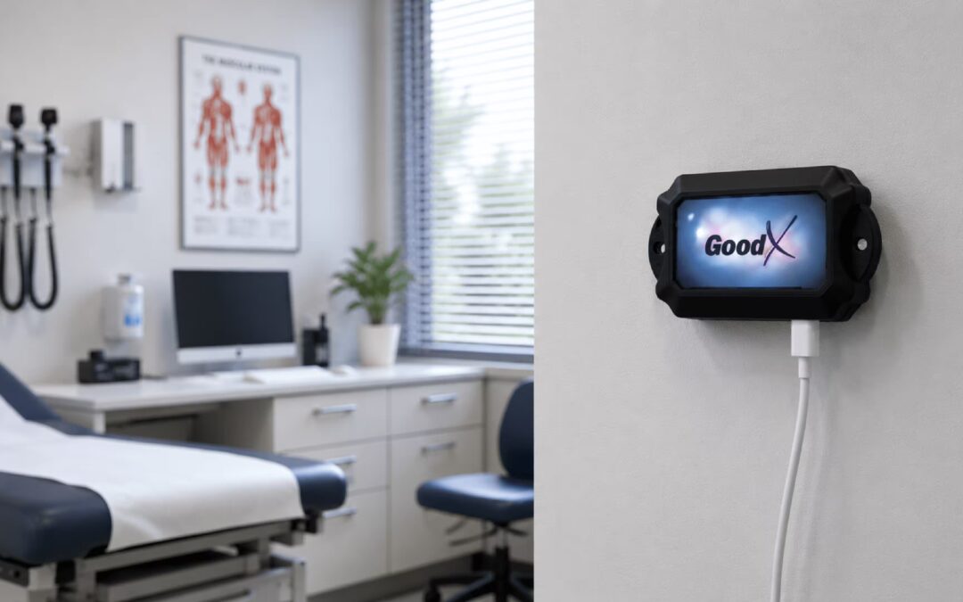
Here is how it works in practice. When a patient arrives at the clinic, they are given an RFID-enabled patient tag or wristband. As the patient moves from the reception area to the pre-testing room, the RFID reader in that zone detects their arrival and automatically updates their status in the GoodX system from “Waiting” to “In Pre-Testing”. When they complete pre-testing and move to the consultation waiting area, the status updates again. When they enter the consulting room, the ophthalmologist’s interface shows the patient is present and their pre-testing results are already attached to their consultation record.
This automatic status advancement eliminates the need for manual check-ins at each stage, reduces the coordination calls between reception, pre-testing technicians, and consulting rooms, and gives the entire clinical team a live view of where every patient is in their visit journey. For a clinic managing 50 to 100 patients a day across multiple consulting rooms and testing stations, this is a significant operational improvement.
The benefits extend to patient experience as well. Patients who can see a clear status display of where they are in their journey, rather than sitting in a waiting room with no information about when they will be seen next, report significantly lower anxiety and higher overall satisfaction with their visit.
Frequently Asked Questions
What is RFID and how is it used in ophthalmology?
RFID (Radio Frequency Identification) is a technology that uses radio waves to identify and track tags without requiring physical contact or line of sight. In ophthalmology, RFID is used for patient flow management, specifically to track patients as they move through the different stages of a clinic visit, from reception through pre-testing to consultation. GoodX uses RFID to advance patient booking status automatically in real time.
Does RFID need line of sight to work?
No. Unlike barcode scanning, RFID does not require line of sight between the tag and the reader. RFID readers can detect tags through walls and around corners, which makes them ideal for clinical settings where patients move between rooms and zones. Detection is automatic and passive, requiring no action from the patient or staff member.
Can RFID be used for patient tracking in an eye clinic?
Yes. RFID is well-suited to patient tracking in an ophthalmology clinic because the multi-step nature of an ophthalmic patient visit creates coordination challenges that manual tracking cannot solve efficiently at scale. GoodX uses RFID specifically to advance patient booking status as patients move through reception, pre-testing, and consultation areas, giving the entire clinical team a real-time view of each patient’s location and progress.
What is the main purpose of RFID in healthcare?
In healthcare, RFID serves several purposes including asset tracking, medication management, and patient flow management. In an ophthalmology clinic context, the primary purpose is patient flow optimisation: tracking where each patient is in their clinic journey and automatically updating their status in the practice management system, so clinical and administrative staff always have an accurate, real-time picture of who is where.
How does RFID improve patient experience in an ophthalmology clinic?
RFID improves patient experience by eliminating manual check-ins at each stage of the visit, reducing unexplained wait times through visible status updates, and enabling the clinical team to coordinate patient movement more efficiently. Patients who can see where they are in their visit journey and who spend less time waiting without information report significantly higher satisfaction scores than those managed through manual coordination systems.
See RFID Patient Tracking in Action with GoodX
GoodX combines advanced ophthalmology practice management tools with emerging technologies including RFID patient tracking to help eye clinics run more efficiently and deliver a better patient experience.
To find out how GoodX can work for your ophthalmology practice, book a free demo with the team at goodxeye.com.

by HannesE | May 27, 2026 | Blogs
Eye care is one of the most documentation-intensive specialties in medicine. Between visual acuity assessments, refraction data, fundus imaging, OCT reports, IOP measurements, surgical planning, and long-term condition monitoring for patients with glaucoma, diabetic retinopathy, or macular disease, the clinical record requirements of an ophthalmology practice are fundamentally different from those of a general medical practice.
Generic practice management software was not built for this. Ophthalmology practice management software that is purpose-designed for eye care handles all of these clinical requirements natively, alongside the administrative workflows that keep an eye clinic running efficiently. This guide covers what ophthalmology practices should expect from their practice management and EMR platform, and how GoodX delivers specifically for this specialty.
Ophthalmology Medical Practice Management: The Full Picture
Ophthalmology medical practice management involves coordinating a significantly more complex set of workflows than a standard GP or general outpatient setting. Consider the appointment structure alone: a single patient visit to an ophthalmology clinic might involve pre-testing by a technician, a refraction assessment, an OCT scan, a consultation with the ophthalmologist, and a follow-up booking all within the same session. Each step has its own documentation, equipment requirements, and staff allocation.
A practice management platform that is designed for ophthalmology understands this structure. It supports multi-step patient journeys within a single visit, connects pre-testing results to the consultation record automatically, and ensures that the complete picture of what happened during the appointment is captured in one place without requiring manual transfer of data between systems or staff members.
Surgical workflow management is another dimension that sets ophthalmology apart. For clinics that perform cataract surgery, LASIK, or other ophthalmic procedures, the practice management system needs to handle theatre booking, pre-operative assessment documentation, IOL calculations, consent forms, post-operative review scheduling, and outcome recording. GoodX Software addresses these workflows as part of a cohesive system rather than as add-on modules bolted to a generic platform.
Financial management for ophthalmology practices also carries specific complexity. Insurance authorisations for certain procedures, billing for diagnostic tests, and managing the mix of medical and optical billing streams requires a billing module that understands this landscape. A general practice billing tool often requires significant manual work-arounds in an ophthalmology context.
According to the World Health Organization, vision impairment affects over 2 billion people globally, and the demand for quality eye care is growing faster than the availability of trained ophthalmologists in most regions. Eye clinics that operate with maximum efficiency through well-designed practice management software are better placed to meet this growing demand without compromising on care quality.
Ophthalmology EMR Software: Clinical Documentation Built for Eye Care
Ophthalmology EMR software needs to do something that general EMR platforms typically cannot: capture the structured clinical data of an eye examination in a format that is medically meaningful, retrievable, and useful for longitudinal patient monitoring.
For a glaucoma patient seen every six months over ten years, the value of the EMR is not just in storing individual visit notes. It is in presenting a structured, comparable view of IOP readings, visual field test results, optic disc assessments, and medication history over time, so the treating ophthalmologist can assess disease progression and treatment response at a glance.
This kind of structured longitudinal data management requires EMR design that goes beyond a generic note-taking interface. Ophthalmology EMR software needs templated examination forms for specific conditions, graphical display of measurements over time, integration with diagnostic equipment outputs, and structured fields for recording standardised clinical parameters.
GoodX is designed with precisely this clinical depth in mind. The platform supports the documentation requirements of an ophthalmology practice from routine refraction through to complex retinal disease management, all within a system that also handles the administrative and billing functions of a busy eye clinic.
The International Agency for the Prevention of Blindness emphasises the importance of comprehensive data collection in eye care settings to support both individual patient outcomes and population-level vision health monitoring. Purpose-built ophthalmology EMR software that captures structured clinical data consistently is a direct enabler of this goal.
Practice Management Software for Ophthalmologists: What GoodXEye Delivers
Practice management software for ophthalmologists needs to solve a specific set of problems that generic platforms handle poorly or not at all. GoodX has been built in response to exactly these challenges.
Speciality-appropriate scheduling handles the unique structure of an ophthalmology clinic day, where appointment types vary significantly in length, the sequence of events within a single patient visit is complex, and equipment availability needs to be coordinated alongside practitioner availability. GoodX Software makes this manageable through a scheduling interface that reflects how ophthalmology clinics actually work.
Diagnostic integration connects the outputs of ophthalmic equipment, including OCT machines, visual field analysers, corneal topographers, and automated refractors, directly to the patient record. When test results flow automatically into the EMR rather than being printed, scanned, and manually attached, the clinical workflow is faster and the record is more complete.
Recall and disease monitoring tools support the long-term patient management that characterises ophthalmology. For patients requiring regular monitoring due to glaucoma, diabetic eye disease, or age-related macular degeneration, automated recall systems ensure follow-up appointments are booked on the appropriate schedule and that patients who are overdue for review are flagged proactively.
According to Health IT Analytics, specialty-specific clinical software consistently outperforms adapted general-purpose platforms on clinician satisfaction metrics, documentation completeness, and workflow efficiency. For ophthalmologists, the difference between a purpose-built platform and a generic EMR is felt on every patient encounter.
Frequently Asked Questions
What is ophthalmology practice management software?
Ophthalmology practice management software is a speciality-specific platform designed to manage the clinical, administrative, and billing workflows of an eye care clinic. It includes structured clinical documentation tools for ophthalmic examinations, appointment scheduling configured for the complexity of ophthalmology clinic visits, diagnostic integration, surgical workflow management, and billing tools that handle the specific requirements of eye care billing.
What should ophthalmology EMR software include?
Ophthalmology EMR software should include templated examination forms for common ophthalmic conditions, structured fields for recording IOP, visual acuity, visual fields, and other standardised measurements, longitudinal data display for monitoring disease progression over time, integration with diagnostic equipment outputs, and clinical documentation tools that support both routine and complex ophthalmic care without requiring work-arounds designed for general practice settings.
Why is GoodX Software a good fit for ophthalmology practices?
GoodX Software is a strong fit for ophthalmology practices because GoodX has been purpose-built for the specific clinical and administrative demands of eye care. It combines specialty-appropriate clinical documentation, complex scheduling support, diagnostic integration, surgical workflow tools, and integrated billing in one platform, rather than requiring ophthalmology practices to adapt a generic system to their specific requirements.
How does ophthalmology practice management software improve patient care?
It improves patient care by ensuring that complete, structured clinical records are available at every encounter, enabling meaningful longitudinal monitoring of conditions like glaucoma and macular disease over time. It also reduces administrative bottlenecks that can delay clinical processes, supports proactive recall management so patients do not miss critical review appointments, and frees clinical staff to focus on patient interaction rather than documentation overhead.
Can ophthalmology practice management software handle surgical scheduling?
Yes. Purpose-built ophthalmology practice management software supports surgical workflow management including theatre booking, pre-operative documentation, IOL calculation records, consent form management, post-operative review scheduling, and outcome recording. This is a key area where specialty-specific software outperforms adapted general practice platforms, which often require manual work-arounds for ophthalmic surgical workflows.
Ready to See GoodXEye in Action?
GoodX is the ophthalmology practice management and EMR platform built specifically for eye care clinics. It combines the clinical depth ophthalmologists need with the administrative and billing tools that keep a practice running efficiently.

by HannesE | May 15, 2026 | Blogs
Ophthalmology billing is among the most code-intensive in all of outpatient medicine. Between the breadth of diagnostic codes covering the full spectrum of eye disease and the procedure codes for both minor and major surgical interventions, getting ICD-10 coding right in an eye care practice requires both a solid grasp of the classification system and the right software tools to apply it efficiently.
This guide is written specifically for ophthalmologists and eye care practice administrators. It covers what ICD-10 diagnosis codes are, how they are constructed, and what you need to understand about their structure to code ophthalmology encounters accurately and compliantly.
What are the ICD-10 Diagnosis Codes?
ICD-10 stands for the International Classification of Diseases, 10th Revision, published by the World Health Organization as the global standard for documenting diseases, health conditions, and clinical encounters. In the United States, the clinical modification of this system, ICD-10-CM, is the version used for outpatient and ambulatory care diagnosis coding and is maintained by the Centers for Medicare and Medicaid Services (CMS) and the National Center for Health Statistics.
For eye care practices, the ICD-10-CM codes that matter most are concentrated in Chapter 7 (Diseases of the Eye and Adnexa), which spans codes H00 through H59. This chapter covers conditions from blepharitis and conjunctivitis to glaucoma, macular degeneration, diabetic retinal disease, and retinal detachments. Eye care practices also regularly use codes from other chapters, including endocrine codes for diabetic eye disease (Chapter 4, E-codes), injury codes for ocular trauma, and Z codes for preventive exams and screening encounters. The World Health Organization ICD portal provides access to the full international classification, while the CDC’s ICD-10-CM resources maintain the US-specific version.
Why Accurate ICD-10 Coding Matters for Ophthalmology
In ophthalmology, diagnostic specificity in ICD-10 coding directly affects medical necessity determinations for both procedures and diagnostic imaging. If a claim for OCT imaging is submitted with an unspecified retinal code when the documentation clearly supports a more specific code, the claim may be denied or downcoded. Similarly, intravitreal injection claims tied to wet AMD require the correct AMD code with the correct laterality and stage to be adjudicated accurately.
The American Academy of Ophthalmology provides specialty-specific coding guidance, including Ophthalmic Coding Coach publications and annual coding updates, that are essential reading for ophthalmology billing staff. Staying current with these resources, alongside the annual ICD-10-CM updates, is part of maintaining a compliant and financially healthy practice.
Anatomy of ICD-10 Codes
Every ICD-10-CM code follows a defined structure, and understanding that structure makes it much easier to navigate the classification system, verify codes, and catch errors before submission. Here is how ophthalmology-relevant codes are built.
Character 1: The Chapter Letter
The first character of every ICD-10-CM code is a letter indicating the broad disease chapter. For diseases of the eye and adnexa, that letter is H. Codes beginning with H00 through H59 cover the full range of ophthalmic conditions from eyelid disorders through retinal diseases. When you see a code beginning with H in an ophthalmology chart, you are in the right chapter. When coding diabetic retinal disease, you will also use E codes (endocrine chapter) as the principal diagnosis, with the H code as a manifestation code.
Characters 2 and 3: The Category
The second and third characters narrow the H-code to a specific disease category. H25 refers to age-related cataract. H26 covers other cataracts. H33 refers to retinal detachments and breaks. H35 is other retinal disorders. H40 is glaucoma. H43 covers the vitreous and H44 disorders of the globe. At this three-character level, you know the general condition but not the specific diagnosis, laterality, or stage.
Characters 4 Through 6: Specificity and Laterality
The fourth through sixth characters, following the decimal point, add essential clinical specificity. In ophthalmology, this is where laterality is captured: right eye, left eye, or bilateral. For example, H40.1110 means: primary open-angle glaucoma (H40.11), mild stage (first .1), right eye (second .1), not specified as low tension or high tension (0). Each additional digit adds a layer of specificity that payers use to adjudicate claims and that the practice record uses to track disease progression over time.
The Seventh Character: Encounter Type
Some ICD-10-CM codes in ophthalmology require a seventh character to indicate the encounter type. Injury codes, in particular, use seventh characters to indicate whether the encounter is initial (A), subsequent (D), or for a sequela (S). An ocular penetrating injury would require the correct seventh character based on where in the course of treatment the patient is being seen. Omitting a required seventh character results in an invalid code and a rejected claim.
Structure of an ICD-10-CM Code
Understanding how the structure of an ICD-10-CM code applies to real ophthalmology encounters helps coders select the right code and avoid the common errors that drive claim rejections in eye care practices.
Laterality: One of the Most Common Ophthalmology Coding Errors
Ophthalmology is one of the few specialties where laterality is embedded in the ICD-10-CM code itself for most conditions. Coding a retinal tear as a unilateral code when the documentation specifies the left eye, or using a bilateral code when only one eye was examined, results in a coding error that can trigger claim rejection or audit. Practice management software with integrated ophthalmology coding support, like GoodX Eye Care, helps prevent laterality errors by guiding coders to the appropriate code level for each documented condition.
Principal and Secondary Diagnosis Sequencing in Ophthalmology
For diabetic eye disease, ICD-10-CM coding guidelines require that the systemic condition, the diabetes code (E10 for type 1, E11 for type 2), be listed as the principal or first-listed diagnosis, with the ophthalmic manifestation code (such as E11.3411 for type 2 diabetes with severe nonproliferative diabetic retinopathy with macular edema, right eye) coded to its full specificity. Getting this sequencing wrong, either by coding the H code first or by using an unspecified diabetes code, is a common error in practices that see high volumes of diabetic patients.
Common Ophthalmology ICD-10-CM Codes Every Practice Should Know
While the full ophthalmology code set is extensive, certain codes appear frequently in most eye care practices. These include the primary open-angle glaucoma codes (H40.11 series with stage and laterality), the age-related macular degeneration codes (H35.31 for dry AMD, H35.32 for wet AMD, both requiring stage and laterality), the diabetic retinopathy codes under E10 and E11, the cataract codes (H25 series for age-related), and refractive error codes (H52 series) used for refraction and spectacle prescription encounters.
Annual Code Updates and Ophthalmology
ICD-10-CM codes are updated each year on October 1. Ophthalmology has been an active area for new and revised codes in recent years, particularly for conditions with evolving treatment protocols such as AMD subtypes and myopia management. CMS publishes the annual ICD-10-CM updates along with a summary of changes. Eye care practices must ensure their coding software and code libraries are updated before October 1 each year to avoid using deleted or invalid codes. GoodX Eye Care applies annual code set updates automatically, eliminating this administrative risk.
Frequently Asked Questions
What are the ICD-10 diagnosis codes used in ophthalmology?
Ophthalmology ICD-10-CM codes are primarily found in Chapter 7 (H00 through H59), covering all diseases of the eye and adnexa. Eye care practices also use Chapter 4 codes for diabetic eye disease, injury codes for ocular trauma, and Z codes for preventive exams and screening encounters. Accurate coding requires selecting codes to the highest level of specificity, including laterality and disease stage.
What is the anatomy of an ophthalmology ICD-10-CM code?
An ophthalmology ICD-10-CM code begins with the letter H (diseases of the eye and adnexa), followed by two digits for the category, a decimal point, and up to four characters specifying the condition, disease stage, and laterality (right eye, left eye, or bilateral). Some injury codes require a seventh character indicating initial, subsequent, or sequela encounter. Each element of the code must match the clinical documentation.
What is the structure of an ICD-10-CM code for eye diseases?
Eye disease codes in ICD-10-CM follow the standard CM structure: a letter indicating the chapter (H for ophthalmology), a two-digit category code, and up to four additional characters that specify the exact condition, stage, and laterality. For example, H35.3210 means: age-related macular degeneration (H35.32), dry type, right eye, stage unspecified. Correct specificity at every level is essential for claim acceptance.
How does laterality affect ICD-10 coding in ophthalmology?
In ophthalmology, most condition-specific ICD-10-CM codes include laterality as part of the code structure. Right eye, left eye, and bilateral codes are often distinct and non-interchangeable. Using the wrong laterality code, even for the correct diagnosis, is a coding error that results in claim rejection or audit risk. Integrated ophthalmology coding software that enforces laterality selection reduces this error type significantly.
How does GoodX Eye Care handle ICD-10 coding for ophthalmology practices?
GoodX Eye Care integrates ophthalmology-specific ICD-10-CM code search directly into the patient encounter. Codes are searched and selected within the clinical record, with built-in guidance for laterality, disease stage, and sequencing requirements for diabetic retinal disease. Annual code set updates are applied automatically, and the coding module connects directly to the billing workflow so claims are built from accurately coded encounter data.
Code Smarter with GoodX Eye Care Software
Accurate ICD-10 coding in an ophthalmology practice is the foundation of a clean revenue cycle. GoodX Eye Care integrates specialty-specific coding directly into the clinical workflow, with automated code updates, laterality guidance, and direct linkage to billing, so your team codes confidently and your claims go out clean. Book a free demo to see how it works.